Low back pain is one of the most common and costly conditions affecting today’s workforce. It is the leading cause of disability in the United States and worldwide, affecting an estimated 80% of adults at some point in their lives, and it ranks as the second most common reason for missed workdays after upper respiratory infections. For workers’ compensation stakeholders, the financial impact is just as significant, with direct and indirect costs estimated at more than $100 billion annually and rising.
For case managers, claims professionals, employers, and rehabilitation specialists, understanding how spine care has evolved is no longer optional. During Medical & Life Care Consulting’s Injury Insight webinar series, The Evolution of Spine Surgery, board-certified orthopedic spine surgeons Marc S. Menkowitz, MD, and Steve J. Paragioudakis, MD, MBA, of the Center for the Functional Restoration of the Spine walked through decades of advancement in lumbar spine care, from early fusion techniques to today’s robotic-assisted, minimally invasive procedures.
Their insights offer a practical roadmap for anyone involved in managing spine-related workers’ compensation claims, helping translate complex surgical advancements into actionable guidance for recovery planning, cost forecasting, and return-to-work strategy.
Understanding the Lumbar Spine and Common Conditions
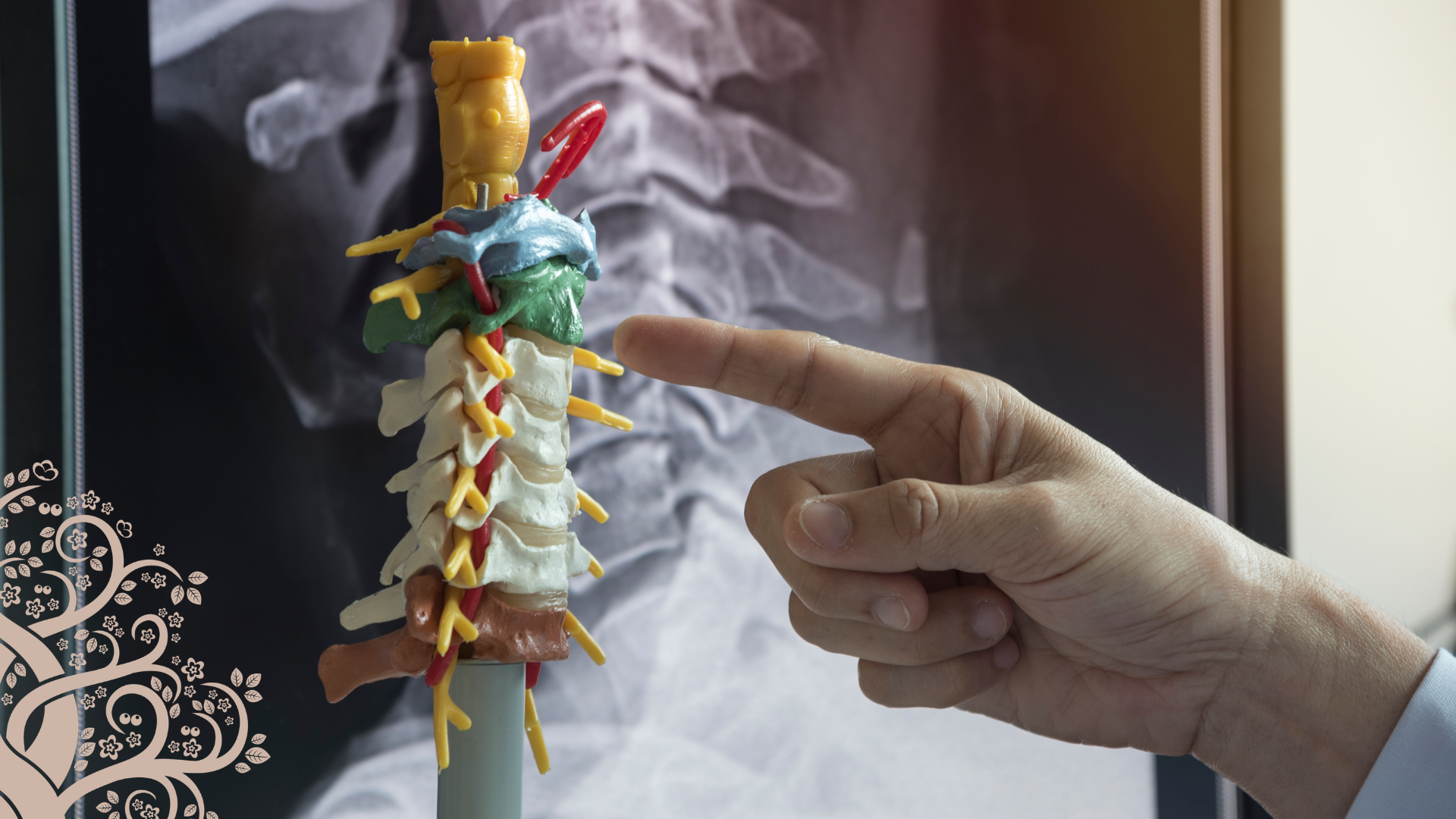
The lumbar spine consists of five vertebrae (L1 through L5) separated by intervertebral discs, with nerves exiting at each level through small openings called neuroforamina. Maintaining the natural curve of the lumbar spine, known as lordosis, is a key surgical goal, since flattening this curve (a complication sometimes seen with older fusion techniques) can create stress on adjacent spinal segments and lead to further degeneration over time.
Several conditions commonly drive workers’ compensation spine claims:
- Disc herniation: Disc material pushes through a tear in the outer wall (annulus) and irritates nearby nerve roots, often causing radiculopathy (sciatica), back pain, or both.
- Spondylolysis: A small stress fracture in part of the vertebra, most often occurring at L5-S1.
- Spondylolisthesis: A forward slip of one vertebra over another, which can be either degenerative (typically at L4-5, developing gradually with age) or isthmic (typically at L5-S1, originating from a pars defect that often begins in adolescence).
- Degenerative disc disease: A natural, progressive loss of disc height and hydration that begins as soon as a person starts sitting upright and can eventually narrow the space available for exiting nerves.
A thorough patient history accounts for the majority of an accurate diagnosis, even before imaging is performed. Diagnostic studies are typically used to confirm a working diagnosis rather than to establish it from scratch, which is an important distinction for claims professionals reviewing the medical necessity of imaging requests.
Most Low Back Pain Resolves Without Surgery
One of the most important takeaways for claims professionals and case managers is this: the overwhelming majority of low back pain cases, more than 90%, are treated successfully without surgery.
Conservative, non-operative care typically progresses through a structured sequence:
- Brief rest (no more than two to three days)
- Anti-inflammatory medication
- Short-term analgesics for acute flare-ups
- Antidepressant medications, which can be effective for radicular pain
- Bracing, when appropriate
- Core-strengthening exercise and activity modification
- Interventional pain management, such as epidural injections, for patients who need a faster path to relief
Surgery is generally reserved for patients who continue to report low back pain rated greater than 6 out of 10 for six months or longer, despite exhausting conservative and interventional options. This threshold is a useful benchmark for claims professionals evaluating treatment timelines and anticipating when a surgical consultation may become appropriate.
The Historical Evolution of Lumbar Fusion
Fusion surgery has undergone several distinct phases of improvement, each one measurably increasing success rates:
- Non-instrumented fusion: Early fusion procedures used bone grafted from the patient’s own iliac crest, laid along the spine without any hardware. These procedures carried a fusion success rate of roughly 72%, with even lower rates of clinical symptom improvement.
- Pedicle screw fixation: The addition of screws and connecting rods provided internal stability, similar to an internal cast, and raised fusion rates to approximately 87%.
- Interbody fusion devices: Modern cage technology, inserted in place of the removed disc and packed with bone graft material, further improved fusion rates to roughly 91%. Interbody devices also restore disc height, help re-establish lumbar lordosis, provide load-sharing that protects the hardware from premature failure, and reduce the risk of pseudoarthrosis (failed fusion).
This progression illustrates why outcomes for patients undergoing fusion today differ meaningfully from those treated decades ago, an important consideration when reviewing older medical records or comparing prior treatment outcomes during case review.
The Shift Toward Minimally Invasive Surgery
Traditional open fusion procedures require stripping muscle away from the spine through a central incision, which contributes significantly to post-operative pain and recovery time. Many practices, including the Center for the Functional Restoration of the Spine, have shifted toward a minimally invasive technique called the MAS T-LIF (minimally invasive transforaminal lumbar interbody fusion), built on a decades-old approach known as the Wiltse approach.
Rather than cutting through the muscle, surgeons work between natural muscle planes through small incisions, sometimes as small as one inch, to perform decompression, remove damaged disc material, and place both screws and an interbody cage.
According to outcomes tracked at the presenting practice, this approach has been associated with:
- Reduced blood loss: from an average of approximately 640 cc with traditional open surgery to approximately 150 cc
- Shorter hospital stays: reduced from an average of 3.2 days to 1.7 days
- Lower opioid use: a 58% reduction in post-operative opioid consumption, as documented through the practice's hospital anesthesia service
- Faster functional recovery: approximately 75% of patients are walking up to one mile without an assistive device within the first week after surgery, with most patients off opioids within 7 to 10 days
There is a learning curve associated with this technique. Surgeons typically need 25 to 50 cases before becoming fully proficient, but the long-term benefits to patients have made it an increasingly standard option for appropriate candidates.
The Role of Robotic-Assisted Navigation
Robotic-assisted spine surgery has become a defining advancement in modern spine care. It’s important to understand what this technology actually is: a navigation system with built-in safeguards, not an autonomous surgical robot. Unlike remote-controlled surgical systems used in other specialties, the surgeon remains scrubbed in and performs every step of the procedure by hand, working through a guided tube, called an end effector, that is attached to the robotic arm.
Before surgery begins, the surgical team obtains an intraoperative CT scan and uses it to pre-plan the precise trajectory for each screw. Tracking markers placed on the patient and the robotic arm continuously communicate with each other; if either one moves unexpectedly, the system halts the procedure until alignment is confirmed. This creates multiple layers of built-in safety checks throughout the case.
Why accuracy matters: Independent studies cited during the presentation found that robotic-assisted screw placement achieves accuracy rates of approximately 98% to 99%, compared to roughly 95% with traditional navigation systems, 90% with live fluoroscopy (X-ray guidance), and 85% with freehand technique based on anatomical landmarks alone. Notably, research also shows that fellows and residents using robotic guidance can achieve accuracy comparable to that of highly experienced surgeons, suggesting the technology helps standardize precision across all experience levels.
The presenting surgeons reported more than seven years of experience with robotic-assisted spine surgery, having performed close to or more than 1,000 procedures and placed upwards of 10,000 screws. As with the minimally invasive technique, there is a learning curve, generally 30 to 50 cases, after which surgeons typically become faster and more precise than they were using freehand methods.
It’s worth noting that robotic navigation does not replace neuromonitoring; the two technologies work in conjunction, with standard neuromonitoring equipment integrated alongside robotic guidance throughout each case.
Expanding What's Surgically Possible
One of the more significant implications of robotic navigation is its ability to safely manage complex revision surgeries that would otherwise carry substantial risk. Examples discussed during the presentation included:
- Placing new screws between existing hardware from a prior, unsuccessful sacroiliac fusion
- Inserting new pedicle screws underneath previously placed hardware to avoid removing an existing construct
- Precisely targeting and removing bone overgrowth that had encroached on a nerve root following a prior fusion
- Adding new instrumentation around old, long-standing hardware without disrupting it
- Adding new instrumentation around old, long-standing hardware without disrupting it
- Treating an isthmic pars fracture with a targeted compression screw, avoiding a fusion altogether in a select, well-suited case
This last example is particularly relevant for claims professionals and case managers evaluating treatment alternatives. A patient in his 30s, a contractor who had been out of work due to a pars fracture and had not improved with pain management or physical therapy, had been recommended for a traditional fusion by multiple surgeons. Using robotic-guided compression screw fixation instead, the fracture was allowed to heal directly. Several years later, the patient had full range of motion, had returned to all normal activities, and had grown his own excavating business. This case illustrates how matching the right technology to the right patient pathology can sometimes provide a less invasive alternative to fusion without compromising long-term outcomes.
What This Means for Recovery and Return-to-Work Planning
For case managers, claims professionals, and employers, these advancements have direct, practical implications:
- Shorter recovery windows are increasingly realistic for well-selected surgical candidates, particularly with minimally invasive and robotic-assisted approaches.
- Reduced opioid reliance post-surgery may simplify medication management and reduce concerns around prolonged opioid use during recovery.
- Faster mobility milestones, such as walking without assistance within the first week, can inform more accurate return-to-work timelines.
- Patient-specific factors still matter significantly. Smoking reduces fusion success rates by roughly half due to its effect on blood flow and tissue healing, and nicotine replacement products (gum, patches) carry the same risk. Obesity also increases surgical complication rates and may affect candidacy for certain procedures, though minimally invasive and robotic techniques can sometimes offer a safer alternative for appropriately selected patients with higher BMI who require decompression.
- Surgical urgency varies by diagnosis. Conditions like spondylolisthesis are rarely surgical emergencies. The decision to proceed depends on symptom severity, duration, and response to conservative treatment, which is useful context when evaluating utilization review requests or treatment delays.
Support Better Recovery Outcomes with Nurse Case Management
Advancements like robotic-assisted spine surgery and minimally invasive fusion techniques offer real promise for injured workers, but technology alone doesn’t guarantee a smooth recovery. Coordinating care across surgeons, physical therapists, pain management specialists, and primary care providers, while keeping employers and claims professionals informed every step of the way, requires dedicated, experienced oversight.
Skilled nurse case management plays a critical role in translating these clinical advancements into real-world results. Effective case management helps:
- Coordinate care among surgeons, specialists, and rehabilitation providers
- Monitor recovery milestones and flag concerns early
- Support injured workers through every phase of treatment, from diagnosis to return to work
- Improve communication among medical providers, employers, and claims professionals
- Facilitate timely access to appropriate treatment, reducing unnecessary delays
- Develop realistic, medically grounded return-to-work plans
- Help manage the complexity of orthopedic and spine-related claims toward cost-effective, sustainable resolution
As spine care continues to evolve, having an experienced nurse case manager involved in complex orthopedic and spine claims can make the difference between a recovery plan that stalls and one that moves an injured worker efficiently toward the best possible outcome.
To learn more about how Medical & Life Care Consulting’s Case Management Services can support your spine-related workers’ compensation claims, we invite you to connect with our team.