Traumatic brain injuries (TBIs) and concussions are complex conditions that can lead to persistent symptoms such as dizziness, headaches, fatigue, cognitive challenges, and balance instability. These issues can affect daily functioning, delay return-to-work timelines, and complicate recovery in workers’ compensation and medical-legal cases, where coordinated care and medical case management often help ensure treatment remains organized and appropriate.
Functional neurology offers a broader perspective on concussion recovery by examining how different brain systems interact and adapt through neuroplasticity. Through detailed diagnostic testing and targeted therapies, clinicians can identify neurological dysfunction and support rehabilitation.
This article is based on insights shared in the Injury Insight webinar, “Practical Applications of Functional Neurology,” presented by Dr. Joshua Helm, DC, DACNB. In this session, Dr. Helm provides an inside look at how functional neurology is used in clinical practice to evaluate and treat patients with traumatic brain injuries (TBI).
Understanding Functional Neurology
Functional neurology is a clinical approach that evaluates how the brain functions as a dynamic system rather than focusing only on structural injury.
After a concussion, patients frequently report a combination of symptoms such as:
• headaches
• dizziness
• vision problems
• fatigue
• anxiety
• difficulty concentrating
While these symptoms may seem unrelated, they often stem from disruptions in how different neurological systems communicate with one another.
Functional neurology identifies these breakdowns by assessing sensory processing, motor control, autonomic regulation, and cognitive integration. By identifying where the brain’s communication networks are struggling, clinicians can design therapies that stimulate specific neural pathways and encourage recovery.
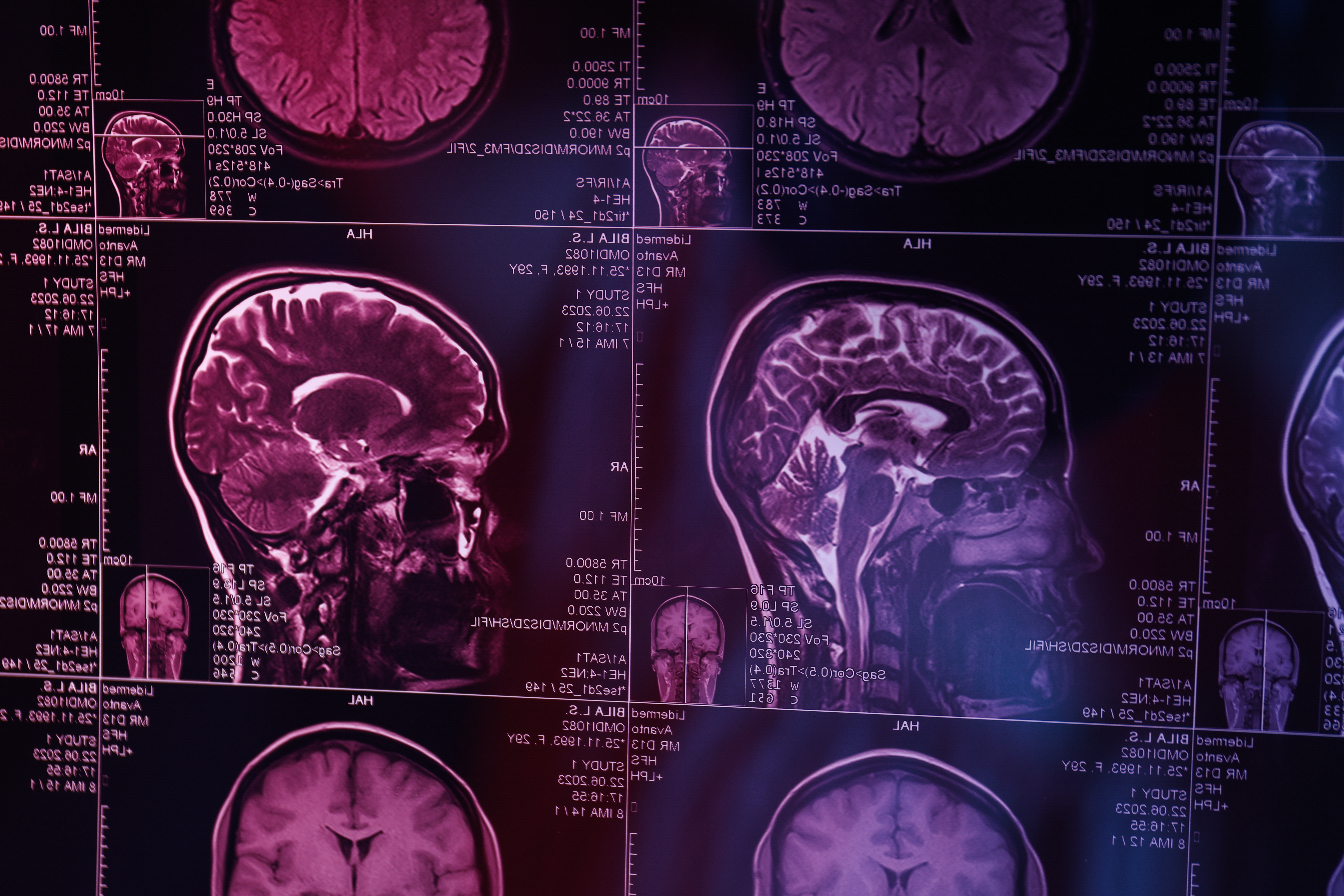
This approach is especially valuable in concussion rehabilitation, where traditional imaging such as CT scans or MRIs may not reveal functional neurological deficits.
The Role of Neuroplasticity in Brain Recovery
A central principle of functional neurology is neuroplasticity, the brain’s natural ability to adapt and reorganize itself.
Neuroplasticity allows the brain to:
• create new neural connections
• strengthen existing pathways
• compensate for damaged areas
• restore communication between brain regions
A common phrase used in neuroscience is:
“Neurons that fire together wire together.”
Repeated stimulation strengthens neural circuits. When specific brain pathways are activated repeatedly through therapy, the brain gradually improves its ability to process information and coordinate bodily functions.
For concussion patients, neuroplastic rehabilitation may include visual training, vestibular stimulation, balance exercises, and coordinated movement therapies designed to activate and reinforce neurological pathways.
How the Brain Interprets the Environment
The brain constantly interprets sensory information to maintain stability and orientation. In functional neurology, this process can be simplified into two fundamental questions the brain continually asks:
Brain Function Question | Purpose |
Where am I in my environment? | Determines spatial orientation |
Where is my environment relative to me? | Helps maintain balance and safety |
To answer these questions accurately, the brain relies on three interconnected sensory systems.
The Three Sensory Systems That Maintain Balance
For the brain to interpret its surroundings correctly, it must integrate information from the visual, vestibular, and proprioceptive systems.
| Sensory System | Function | Symptoms When Disrupted |
| Visual System | Processes movement and environmental cues | blurred vision, eye strain |
| Vestibular System | Located in the inner ear; controls balance and motion detection | dizziness, vertigo |
| Proprioceptive System | Detects body position through muscles and joints | poor coordination |
When these systems provide consistent information, the brain maintains stable balance and spatial awareness.
However, when one system provides conflicting signals, the brain experiences a sensory mismatch. This mismatch can produce symptoms commonly associated with concussion, including dizziness, motion sensitivity, fatigue, and anxiety.
Functional neurological rehabilitation aims to restore communication between these systems so that the brain can interpret sensory information accurately.
Why the Brainstem Is Important in Rehabilitation
Many rehabilitation strategies focus on the brainstem, a region responsible for regulating essential neurological functions.
The brainstem controls several processes that directly affect concussion symptoms, including:
• eye movement coordination
• balance reflexes
• heart rate regulation
• blood pressure control
• breathing patterns
• autonomic nervous system activity
Because the brainstem integrates signals from the visual and vestibular systems, dysfunction in this region can produce widespread symptoms.
| Brainstem Function | Possible Symptoms |
| Vestibular integration | dizziness and imbalance |
| Ocular motor control | blurred or unstable vision |
| Autonomic regulation | fatigue, anxiety |
| Blood flow regulation | headaches |
Rehabilitation therapies that stimulate brainstem pathways can therefore produce improvements across multiple symptoms simultaneously.
Diagnostic Tools Used in Functional Neurology
Before beginning treatment, clinicians conduct comprehensive neurological evaluations that examine how the brain processes sensory information.
Several diagnostic technologies are commonly used in functional neurology clinics.
Computerized Posturography
Computerized posturography is widely considered one of the most reliable tools for measuring balance and sensory integration.
During testing, patients stand on a platform that records body sway while conditions change.
Testing conditions may include:
• standing with eyes open or closed
• turning the head in different directions
• standing on stable or unstable surfaces
The results reveal how effectively the visual, vestibular, and proprioceptive systems work together to maintain balance.
Because the test produces objective data, clinicians can track improvements throughout rehabilitation.
Video Nystagmography
Video nystagmography (VNG) is used to analyze eye movements and vestibular function.
Using infrared cameras in a dark environment, clinicians observe:
• eye movement speed
• eye alignment
• reflex responses
• vestibular-ocular reflex function
The vestibular-ocular reflex (VOR) stabilizes vision when the head moves. If this reflex is impaired, patients may experience blurred vision during movement, dizziness, or difficulty focusing.
Identifying these abnormalities helps clinicians design targeted exercises that retrain visual and vestibular coordination.
Interactive Metronome Therapy
Another tool used in functional neurological rehabilitation is interactive metronome therapy, which focuses on improving timing and coordination between brain regions.
During these exercises, patients perform movements synchronized with a rhythmic auditory beat.
This therapy helps improve:
• motor coordination
• reaction time
• attention and focus
• cognitive processing
Interactive metronome training has been used in concussion rehabilitation as well as in conditions such as ADHD, Parkinson’s disease, and autism spectrum disorders.
Case Study: Autonomic Dysfunction After Concussion
One patient, a 28-year-old woman, presented with persistent symptoms following a concussion. Her symptoms included headaches, dizziness, nausea, and delayed fatigue after physical activity.
She also had Postural Orthostatic Tachycardia Syndrome (POTS), a condition that causes rapid increases in heart rate when standing.
Testing revealed that her visual system struggled to stabilize during movement. This instability disrupted her autonomic nervous system, leading to increased heart rate and dizziness.
Through targeted vestibular and visual stabilization exercises, clinicians improved her gaze stability. As her visual system began functioning more effectively, her autonomic symptoms also improved.
This case illustrates how visual dysfunction can influence broader physiological systems.
Case Study: Mast Cell Activation Syndrome and Balance Instability
Another patient presented with Mast Cell Activation Syndrome (MCAS), a condition that causes heightened sensitivity to environmental triggers.
Patients with MCAS may experience symptoms such as inflammation, headaches, dizziness, and extreme reactions to environmental stimuli.
Balance testing showed that the patient’s stability worsened significantly when her eyes were closed, suggesting vestibular dysfunction.
Treatment focused on regulating the autonomic nervous system through breathing techniques and vestibular rehabilitation.
Follow-up testing demonstrated improved balance and reduced sway patterns.
Case Study: Vestibular Injury from Water Skiing Accident
A 62-year-old patient sustained a ruptured eardrum during a high-speed water skiing accident.
Balance testing revealed severe instability when the patient tilted his head backward. This position disrupted signals from the injured inner ear, causing spatial disorientation.
Rehabilitation focused on retraining the vestibular system using controlled head movements and balance exercises.
Within two weeks, the patient showed measurable improvements in balance testing and reported reduced dizziness.
Why Individualized Treatment Plans Matter
One of the most important principles in functional neurology is that each brain injury is unique.
Two patients with similar concussions may experience completely different symptoms depending on their medical history, neurological function, and environmental factors.
Effective rehabilitation therefore requires:
• detailed neurological evaluation
• individualized therapy strategies
• ongoing monitoring of progress
Standardized treatment plans may overlook the specific neurological dysfunction responsible for symptoms.
Functional neurology instead focuses on identifying and treating the exact neurological mechanism underlying a patient’s condition.
Intensive Rehabilitation and Treatment Duration
Functional neurological rehabilitation often follows an intensive treatment model designed to maximize neuroplastic adaptation.
| Treatment Model | Duration |
| Neuro-intensive rehabilitation | 2 hours per day for 1 week |
| Modified intensive rehabilitation | 1 hour per day for 2 weeks |
Frequent therapy sessions provide repeated neural stimulation, encouraging the brain to reorganize and strengthen functional pathways.
Patients are usually given home exercises following intensive treatment to maintain and reinforce progress.
Functional Neurology in Traumatic Brain Injury Rehabilitation: Final Insights
Traumatic brain injuries often affect multiple neurological systems, making diagnosis and rehabilitation complex. While traditional imaging may not always reveal functional disruptions, approaches such as functional neurology help clinicians better understand how sensory integration, motor control, and autonomic regulation influence recovery.
In complex injury cases, coordinated clinical oversight can play an important role in ensuring treatment remains aligned with a patient’s evolving needs. Through medical case management services, care can be organized across providers, rehabilitation progress monitored, and treatment strategies supported throughout the recovery process.