A worker is injured in an ATV rollover. He can move his hand, but his shoulder and elbow are completely paralyzed. He’s told by several providers to wait and see. Eight months later, he finally reaches a specialist. Still inside the window for meaningful reconstruction, but just barely.
For adjusters managing reserves and case managers coordinating care, that gap has real consequences: higher long-term costs, slower recovery, and in some cases, permanent impairment that could have been reduced.
This post is based on a presentation by Dr. Rohit Garg, fellowship-trained hand and upper extremity surgeon at Mass General Brigham, delivered during the MLCC Injury Insight webinar series. Watch the full session here: Brachial Plexus and Peripheral Nerve Injuries — MLCC Injury Insight.
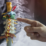
What Is the Brachial Plexus and Why Do These Claims Look Different?
The brachial plexus is a network of nerves running from the spinal cord (levels C5–T1) through the neck and into the arm. It controls all movement and sensation in the shoulder, arm, forearm, and hand. When these nerves are damaged, the results can be severe: loss of shoulder or elbow function, numbness, and chronic neuropathic pain.
These are not soft-tissue strains. They are structural nerve injuries that frequently require surgery, intensive rehabilitation, and recovery timelines measured in years, not weeks. They also rarely occur in isolation.
Common Associated Injuries
Because the mechanism is almost always high-energy trauma, brachial plexus injuries typically present alongside:
- Rib fractures and pneumothorax
- Scapular or clavicle fractures
- Cervical spine fractures
- Vascular injuries (the subclavian artery runs directly alongside these nerves)
- Traumatic brain injury or concussion
Each of these comorbidities adds treatment complexity and affects the sequencing of care. Adjusters should expect multi-specialty involvement from the outset.
How These Injuries Happen
The typical mechanism is the neck and shoulder being pulled in opposite directions, stretching or tearing the nerve roots. Common causes in workers’ compensation include motorcycle and ATV accidents, industrial equipment incidents, falls from height, and crush injuries.
Injury Severity: Why Classification Matters for Reserves and Prognosis
Not every brachial plexus injury has the same prognosis or cost profile. The key variable is how severely the nerve was damaged and at what leve
Three Injury Grades
In neurapraxia, the nerve’s conducting unit (the axon) is intact—only the outer sheath is affected. These injuries recover on their own, typically within weeks to a few months, and carry the best prognosis. In axonotmesis, the axons are disrupted internally. The nerve must regenerate at approximately one millimeter per day. Depending on how far the nerve needs to grow to reach the target muscle, this process can take many months. In neurotmesis, the nerve is completely severed. Without surgical reconstruction, meaningful recovery is unlikely.
Avulsion vs. Rupture: The Distinction That Changes Everything
Beyond injury grade, there is a critical anatomical distinction that shapes the entire treatment and prognosis picture. In a root avulsion, the nerve root is torn off the spinal cord itself—direct repair is not possible. In a rupture, the nerve root remains attached to the spinal cord but is torn at a point further down, which allows for reconstruction.
Avulsion injuries represent the most severe end of the spectrum. When imaging confirms root avulsion, the affected nerve root will not recover regardless of intervention. Life care plans and reserve projections for avulsion-level injuries should reflect permanent impairment from the outset.
The 18-Month Window Every Case Manager Should Know
Muscle motor end plates, the connection points where nerves activate muscles, survive for approximately 18 months after losing their nerve supply. After that threshold, those muscles cannot recover even if the nerve regenerates or surgery is performed. This creates a hard deadline that has direct implications for authorization and referral decisions.
A delayed authorization for specialist evaluation or surgical consultation is not a minor administrative inconvenience in these cases. It can directly determine whether an injured worker recovers functional use of their arm or does not.
Diagnosis and Workup: What Studies to Expect and When
A complete workup for brachial plexus injury involves multiple studies. Understanding the purpose and timing of each helps case managers support appropriate authorization and avoid gaps in documentation.
Clinical Examination
A detailed manual muscle test by a brachial plexus specialist maps exactly which nerve roots are functioning, identifies atrophy patterns, and distinguishes injury level. This examination is the foundation of the treatment plan and requires a specialist with specific expertise, not a general orthopedist or emergency physician.
Imaging
Plain X-rays of the cervical spine, chest, and shoulder establish baseline bony injury. MRI of the brachial plexus (using specialized sequences) is the preferred study for visualizing nerve root status. CT myelogram is an alternative. Both are most informative at three to four weeks post-injury, when pseudomeningoceles (fluid pockets that form when nerve roots are torn from the spinal cord) have had time to develop. Imaging requested in the first days after injury may significantly underestimate the extent of damage.
EMG and Nerve Conduction Studies
Electrodiagnostic testing (EMG/NCS) quantifies the degree of denervation and tracks recovery over time. The optimal window is four to six weeks post-injury. Earlier studies will not yet reflect the full extent of nerve damage due to the time required for Wallerian degeneration (axon breakdown) to complete.
Case managers should expect EMG to be repeated at intervals throughout recovery. Serial studies provide objective evidence of whether nerve regeneration is occurring and whether the treatment plan is on track.
Surgical Options and What They Mean for Recovery Timelines
Treatment selection depends on injury type, time since injury, and which nerve structures remain available. Dr. Garg’s webinar walked through three clinical cases that illustrate the range of approaches.
Nerve Transfer
For post-ganglionic ruptures (where the nerve root is still attached to the spinal cord), nerve transfer is the preferred surgical approach. The surgeon harvests an “expendable” fascicle from a nearby functioning nerve and connects it directly to the non-functioning nerve close to the target muscle. Because the transfer is performed near the muscle, the nerve has a shorter distance to regenerate—improving recovery speed and outcomes.
Dr. Garg’s published research found nerve transfer outcomes were superior to nerve grafting for complete traumatic upper plexus injuries. In one patient, biceps activation was visible at three months post-surgery.
Neurolysis (Scar Release)
When a nerve is structurally intact but compressed by scar tissue, surgical release can restore function without reconstruction. This is a less invasive procedure with a more favorable recovery profile. It requires precise pre-operative identification through imaging and electrodiagnostic testing. It is frequently overlooked at non-specialist centers.
Nerve Grafting
For avulsion injuries or complete plexus injuries where roots are unavailable, a donor nerve (typically from the leg) is used to bridge the gap. Recovery timelines are longer than nerve transfer because the regenerating nerve must travel a greater distance.
Free Functioning Muscle Transfer and Amputation
For the most severe complete avulsion cases, free functioning muscle transfer (using the gracilis muscle from the thigh, transplanted with its nerve and blood supply) can restore limited but meaningful function. Amputation, while rarely first-line, is a legitimate option in select cases and can be combined with targeted muscle reinnervation to reduce phantom limb pain and improve prosthetic function.
What Case Managers Need to Coordinate
Brachial plexus injuries require true multidisciplinary management—not sequential referrals, but coordinated team-based care. The Mass General Brigham Brachial Plexus Program integrates hand surgeons, plastic surgeons, neurophysiologists, occupational and physical therapists, and pain specialists operating as a single program. For case managers supporting injured workers outside major academic centers, replicating this coordination is the central challenge. MLCC’s case management services support this coordination across complex injury cases.
Pre-Operative Occupational Therapy
OT begins before surgery. Pre-operative therapy strengthens the muscle groups that will serve as nerve donors and maintains joint mobility. Stiff joints will not respond to nerve recovery regardless of how successful the surgery is. Range-of-motion preservation is a clinical priority from day one.
Post-Operative Motor Relearning
After nerve transfer surgery, patients must learn to generate a new mental command to activate a transferred nerve’s new target. For example, a patient whose biceps is now powered by a nerve previously controlling wrist flexion must think “move my wrist” to bend their elbow. This cognitive relearning is intensive, highly specific, and cannot be replicated with generic upper extremity therapy protocols. Authorizing a therapist with nerve transfer experience is essential.
Mirror therapy and EMG biofeedback support motor relearning and double as pain management tools. These modalities should be included in the treatment plan and authorization from early in the post-operative phase.
Pain Management
Neuropathic pain is a major comorbidity in brachial plexus injuries and can persist long after structural recovery. Medications commonly used include Gabapentin, Lyrica (pregabalin), and SSRIs. Pain specialists should be part of the care team, not a late-stage add-on. CRPS Type 2 is a recognized secondary condition in nerve injury cases. Case managers should monitor for its development and ensure the treatment plan addresses it proactively.
Psychosocial Support
Dr. Garg noted that social work is often the missing component in brachial plexus programs. These injuries are life-altering. Loss of dominant hand function, inability to return to prior occupation, prolonged disability, and severe pain create significant psychological burden. Counseling and psychological support are clinically indicated and should be built into the care plan, not treated as optional.
Reserve-Setting Benchmarks and Return-to-Work Considerations
Claims reserves for brachial plexus injuries should reflect realistic timelines. Compressed timelines based on soft-tissue injury norms will consistently underestimate costs.
Recovery Timeline by Phase
- 0–4 weeks post-surgery: immobilization; sling use; no active therapy
- 4–8 weeks: gentle passive range-of-motion; possible introduction of e-stim
- 2–3 months: active OT begins; motor relearning initiated for nerve transfer patients
- 6–12 months: first meaningful assessment of surgical outcomes; functional gains emerging
- 12–24 months: continued neurological recovery; maximum medical improvement typically not before this point
- Beyond 24 months: residual deficits are likely permanent; long-term accommodation and vocational planning apply
Return-to-Work Realities
Return to prior occupation, particularly manual labor, construction, or trades, is frequently not achievable after significant brachial plexus injury. Early vocational assessment is appropriate for cases involving dominant extremity injury or complete plexus palsy. Modified duty options should be explored in parallel with rehabilitation, but physical demands must be realistically calibrated to the injury level and recovery phase.
For additional resources on complex injury management and care coordination, visit the MLCC Insights page.
Key Takeaways
- Brachial plexus injuries are high-energy, multi-structure injuries with recovery timelines of 12–24 months—reserves set on soft-tissue norms will fall short
- The 18-month muscle end plate window is a hard biological deadline—authorization delays that push specialist evaluation past this window can permanently limit recovery
- Injury classification (neurapraxia vs. axonotmesis vs. neurotmesis; avulsion vs. rupture) determines prognosis and cost profile—case managers should understand which type is documented
- EMG/NCS should be obtained at four to six weeks post-injury, not immediately—early studies underestimate severity
- MRI of the brachial plexus is the appropriate imaging study; standard shoulder or neck MRI is insufficient
- Specialist referral should go to a brachial plexus program or fellowship-trained hand surgeon—not a general orthopedist
- OT with nerve transfer expertise is essential post-operatively; generic upper extremity therapy protocols are not appropriate
- Pain management, including CRPS monitoring, and psychosocial support are clinical necessities, not optional add-ons
Frequently Asked Questions
What kind of specialist should a brachial plexus injury be referred to?
Fellowship-trained hand surgeons with specific brachial plexus expertise, or neurosurgeons at dedicated brachial plexus programs. General orthopedic surgeons or shoulder specialists without this training are not appropriate primary referrals for complex cases. Centers such as Mass General Brigham, HSS in New York, and Mayo Clinic operate dedicated multidisciplinary programs.
How soon should the referral happen?
As soon as possible after the acute phase is stabilized—ideally within the first four to six weeks. Given that muscle end plates survive for only 18 months post-injury, any delay in reaching a specialist compresses the window for surgical intervention. Research shows nearly half of patients experience referral delays of more than three months, with measurable impact on outcomes.
Why is the EMG ordered weeks after the injury rather than immediately?
Because the nerve degeneration process that EMG measures—Wallerian degeneration—takes three to four weeks to complete after injury. Studies obtained in the first week or two post-injury may show near-normal results even in severe injuries, leading to underestimation of damage. The optimal window for meaningful electrodiagnostic testing is four to six weeks post-injury.
What is CRPS and how does it relate to brachial plexus injuries?
Complex Regional Pain Syndrome (CRPS) Type 2 is a condition in which nerve injury triggers a cascade of autonomic dysregulation—resulting in severe pain, temperature and color changes, and hypersensitivity. It is a recognized secondary complication of peripheral nerve injuries, including brachial plexus injuries. Management is multidisciplinary and may include Gabapentin, Lyrica, mirror therapy, e-stim, and pain specialist intervention. Case managers should monitor for CRPS symptoms and ensure the treatment plan addresses them.
When is it appropriate to consider amputation?
Amputation is considered in complete avulsion injuries where reconstruction has not restored meaningful function, or where a flail, insensate limb poses a safety risk or functional disadvantage compared to a prosthetic. When nerve stumps are available, targeted muscle reinnervation (TMR) or regenerative peripheral nerve interface (RPNI) techniques are used at the time of amputation to reduce phantom limb pain and improve prosthetic control. This decision requires specialist input and should not be made without evaluation at a brachial plexus center.
Conclusion
Brachial plexus injuries are among the most complex and costly cases that adjusters and case managers encounter. The clinical pathway is time-sensitive, multi-disciplinary, and does not fit standard soft-tissue claims management models. Understanding the 18-month window, the importance of specialist referral, and what realistic recovery looks like enables better authorization decisions, more accurate reserves, and meaningfully better outcomes for injured workers.
MLCC’s certified nurse case managers and life care planners support claims teams navigating complex injury cases from acute management through long-term planning. Learn more at medicalandlifecare.com/insights or connect with our team through our case management services page.